
Stroke: Hidden Dangers and Critical Warning Signs You Must Know, Part 1
A stroke is commonly imagined as a sudden catastrophe with obvious symptoms: paralysis, speech impairment, or loss of consciousness. In reality, however, the condition does not always develop so noticeably. A person may continue working, talking, and going about their usual activities without realizing how serious their condition is.
What makes a stroke particularly dangerous is that it can affect not only movement and speech, but also a person’s ability to correctly assess what is happening, make decisions, and seek help independently.
Why can even doctors, who are well aware of the signs of stroke, fail to recognize the danger in time? And what role do people around them play in saving their lives?
Two real-life stories help answer these questions - those of well-known American dermatologist Sandra Lee and neuroanatomist Jill Bolte Taylor.
Dr. Sandra Lee: The Stroke That Nearly Ended a Surgeon’s Career
Dr. Sandra Lee is an American dermatologist and dermatologic surgeon who became widely known for videos of cyst, lipoma, and other skin growth removals. Her YouTube channel, Dr. Pimple Popper, attracted millions of followers. In 2018, her own television show premiered on TLC, and later, Lifetime introduced Dr. Pimple Popper: Breaking Out.
A television crew is constantly present during filming at her clinic, so Dr. Lee’s working day involves not only consultations and surgical procedures but also numerous organizational responsibilities.
On November 20, 2025, while filming a television program, Dr. Lee, who was 55 years old at the time, suddenly felt an intense sensation of heat and began sweating. Initially, she thought it was an ordinary menopausal hot flash and continued working.
That evening, Dr. Lee went to her parents’ home, located near the clinic. She felt anxious, could not sleep properly, and experienced shooting pain in her leg. During the night, while trying to walk downstairs, she discovered that walking had become difficult.
The following morning, weakness on the left side of her body became more noticeable. Dr. Lee performed a simple test herself: she extended both arms in front of her and tried to keep them at the same level. Her left arm gradually drifted downward. Her speech had also changed - her words sounded unusually slurred. The combination of arm weakness, speech difficulties, and walking problems provided strong grounds to suspect a stroke.
However, Dr. Lee continued looking for other explanations. She considered fatigue, anxiety, lack of sleep, and her demanding work schedule. She was concerned about patients who had traveled for surgery and the scheduled filming. Despite her own suspicions, she still managed to style her hair, apply makeup, and put on her medical uniform.
Her parents eventually convinced her to go to the hospital. Dr. Lee’s father was also a dermatologist and understood the danger of the symptoms she was experiencing.
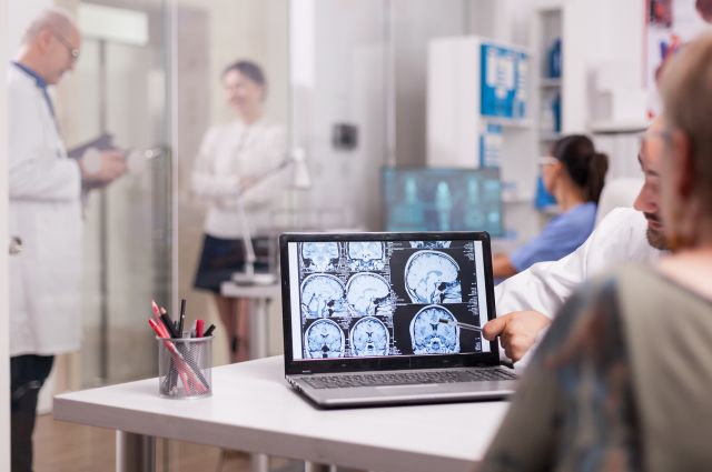
In the emergency department, magnetic resonance imaging (MRI) confirmed an ischemic stroke. However, too much time had already passed since the first symptoms appeared for her to receive standard treatment with a clot-dissolving medication.
For Dr. Lee, the diagnosis was particularly devastating because of its professional consequences. She noticed that her left hand had become weaker and that her movements had lost their previous precision. The work of a dermatologic surgeon requires constant control of fine finger movements in both hands, and even relatively minor impairments can make surgical procedures unsafe for patients.
Filming had to be suspended for approximately two months. Dr. Lee underwent physiotherapy and movement rehabilitation, retraining her balance and the precision of her hand movements. To keep the clinic operating, she invited her longtime friend and colleague, dermatologic surgeon Dr. Kristen Townley, whom she had known since their specialty training.
In January 2026, Dr. Lee gradually returned to practice, although she was initially concerned about performing complex surgical procedures. In an interview with People in April, she explained that she had largely recovered but continued to notice changes in her speech and some movement limitations, particularly when overtired.
Looking back, Dr. Lee acknowledged that she had not adequately monitored her blood pressure and cholesterol levels. She also said that she had underestimated her symptoms, even though she knew how dangerous delaying medical attention could be.
What Happens to the Brain During a Stroke
The brain consumes significant amounts of oxygen and glucose but has virtually no energy reserves that would allow it to function for long without a continuous blood supply. Therefore, disrupted blood flow affects nerve cell function relatively quickly.
An ischemic stroke, which is what Dr. Lee experienced, develops when a cerebral artery becomes blocked. A blood clot - a thrombus - may form directly inside a vessel affected by atherosclerosis or travel to the brain through the bloodstream from another part of the body, such as the heart.
However, an ischemic stroke can also occur when small arteries located deep within the brain are affected. These vessels are particularly sensitive to long-term high blood pressure and metabolic disorders, especially diabetes mellitus. Their walls gradually undergo changes, the lumen narrows, and when a blockage occurs, the blood supply to a small area of the brain is interrupted. This type of stroke is called a lacunar stroke.
Despite the small size of the damaged area, a lacunar stroke can cause significant neurological impairments. Its symptoms depend on the location of the affected region. For example, sudden weakness or numbness on one side of the body, impaired coordination, clumsiness of hand movements, or slurred speech may occur. At the same time, difficulties understanding speech, visual field loss, and other signs of damage to the cerebral cortex are less characteristic of a classic lacunar stroke.
When blood flow stops, nerve cells in the most severely affected area quickly begin to die. However, an area of brain tissue often remains around it where blood flow is significantly reduced but the cells are still alive. If blood flow can be restored in time, some of this tissue may be saved.
This is why it is so important to remove or dissolve the clot quickly during an ischemic stroke. The sooner blood begins flowing again to the affected area, the less extensive the brain damage may be.
But a stroke is not always caused by a blocked vessel. During a hemorrhagic stroke, a blood vessel ruptures and bleeding occurs. The blood damages surrounding nerve cells, while the accumulated blood can compress neighboring areas of the brain.
One of the main causes of this type of bleeding is persistently elevated blood pressure, which gradually damages the walls of small blood vessels. However, bleeding can also result from congenital abnormalities of blood vessel structure, age-related changes in vessel walls, diseases affecting blood clotting, and other causes.
A ruptured aneurysm is particularly dangerous. An aneurysm is a weakened section of an artery wall that forms a bulge. When it ruptures, blood may enter the space between the brain and the membranes surrounding it. This type of bleeding is often accompanied by a sudden, extremely severe headache.
Ischemic and hemorrhagic strokes require fundamentally different treatments. Medications that dissolve blood clots may help when an artery is blocked, but during bleeding they can significantly worsen the hemorrhage.
At the same time, the symptoms of both types of stroke can be very similar. Both may cause sudden limb weakness, speech difficulties, impaired coordination, and altered consciousness. Therefore, external symptoms alone cannot reliably determine whether a blood vessel has become blocked or bleeding has occurred.
The story of neuroanatomist Dr. Jill Bolte Taylor, who experienced a brain hemorrhage, helps illustrate how differently a person may behave during a stroke.
Dr. Jill Bolte Taylor: The Neuroanatomist Who Observed Her Own Brain Hemorrhage
On December 10, 1996, Dr. Jill Bolte Taylor, a 37-year-old American brain researcher at Harvard Medical School, woke up with an unusual throbbing pain behind her left eye.
A hemorrhage caused by the rupture of an arteriovenous malformation - a congenital abnormal connection between arteries and veins - had occurred in her left cerebral hemisphere.
Initially, Dr. Taylor tried to continue her usual morning routine. However, while exercising on a machine, she noticed that her movements felt strange and that her own body seemed unfamiliar. Later, while in the bathroom, she experienced problems with balance, and her movements became slow and stiff.
As the bleeding progressed, her perception of the surrounding world also changed. Dr. Taylor described how her usual internal dialogue gradually disappeared. She began losing the clear sense of the boundaries of her own body and periodically entered an unusual state of calm and euphoria. When her right arm became completely paralyzed, the researcher realized that she was having a stroke.
However, even recognizing what was happening did not lead her to seek help immediately. Dr. Taylor continued to periodically lose touch with her surroundings, while thoughts of danger gave way to a feeling of extraordinary peace.
She then faced a task that would normally have required no effort: making a telephone call. She decided to contact her colleagues at work but could not remember the number. Knowing that it was printed on a business card, Dr. Taylor took out an entire stack of cards and began looking for the correct one.
However, letters and numbers had already lost their meaning for her. She could see individual lines, shapes, and images but could no longer read what was written. Dr. Taylor later recalled that she had managed to go through only a small part of the stack of business cards in 45 minutes. Throughout this time, her condition continued to deteriorate.
When she finally found the correct card, an even more difficult problem arose. She could not understand numbers or use the telephone in the usual way. Instead of reading the number and dialing it, Dr. Taylor began matching the shapes of the symbols on the card with the numbers displayed on the telephone buttons. She periodically forgot which buttons she had already pressed, so she used her paralyzed right hand to cover the numbers she had already dialed.
Later, people asked why she had not called 911 directly. Dr. Taylor explained that, because of the brain damage, numbers had lost their usual meaning. The possibility of dialing 911 simply did not occur to her as an available way to obtain help. The only plan she could still hold in her mind was calling her workplace.
Eventually, she managed to reach a colleague. But when he answered, Dr. Taylor could not understand a single word. His speech sounded like meaningless noises. She tried to identify herself and ask for help. In her mind, the sentence sounded clear, but only unintelligible sounds came out.
Fortunately, her colleague realized that something serious had happened and arranged for help. Dr. Taylor was taken to a hospital and later transferred to Massachusetts General Hospital. On December 27, neurosurgeons removed a large blood clot approximately the size of a golf ball that was compressing areas of the brain associated with speech.
Her recovery took approximately eight years. Dr. Taylor had to relearn reading, writing, speaking, and many everyday activities. Her mother played an enormous role in her rehabilitation.
In 2008, Dr. Taylor published the book My Stroke of Insight: A Brain Scientist’s Personal Journey and delivered a TED talk. The account of a researcher who was able to describe the gradual loss of her own neurological functions became one of the most widely known presentations about stroke.
The stories of these two doctors illustrate different reasons for dangerous delays in seeking help. Dr. Lee retained the ability to reason and make decisions but postponed going to the hospital. Dr. Taylor understood that she was having a stroke but gradually lost the ability to read, speak, and perform sequential actions. In both cases, the people around them played a decisive role in obtaining medical assistance.
Why Recognizing a Stroke in Time Is So Important
According to estimates from the Canadian organization Heart & Stroke, more than 108,000 strokes occur in the country each year - approximately one every five minutes. Nearly one million Canadians live with the consequences of a previous stroke.
According to Statistics Canada, 13,680 people died from cerebrovascular diseases, including stroke and its consequences, in 2024. This group of diseases ranked fourth among the leading causes of death in Canada.
Although the risk of stroke increases sharply with age, it cannot be considered a disease affecting only older adults. According to Public Health Agency of Canada data for 2023-2024, the annual incidence of new cases was approximately 261 per 100,000 people aged 50-64, 675 among those aged 65-79, and 1,664 among people over 80.
Particularly interesting findings came from British Columbia. In a study published in the Canadian Journal of Neurological Sciences in November 2025, researchers analyzed 123,075 stroke events between 2002 and 2022.
Overall, stroke incidence was declining, but the opposite trend was observed among residents aged 35-54: an increase of 14% among women and 27% among men.
Men generally have a higher risk of stroke at comparable ages, but women tend to experience strokes at older ages. In the BC study, the average age at stroke was 79 years for women and 74 years for men.
A Heart & Stroke survey published in June 2025 found that more than one-third of Canadians do not know the most characteristic symptoms of stroke. Despite how common the disease is, public awareness of its signs remains insufficient.
How to Recognize a Stroke
In Canada, the abbreviation F.A.S.T.: is used to remember the most characteristic signs of stroke:
-
Face - sudden facial asymmetry: one corner of the mouth droops, and the smile becomes uneven;
-
Arms - unexpected weakness in one arm: the person cannot raise both arms or keep them at the same level;
-
Speech - the person suddenly begins speaking unclearly, struggles to find words, or stops understanding what others are saying;
-
Time - if any of these signs appear, call 911 immediately.

However, the F.A.S.T. system does not cover all possible manifestations of stroke.
Sometimes the first signs are sudden dizziness, an unsteady gait, loss of balance, or an inability to perform familiar movements. A person may unexpectedly begin bumping into objects, miss an object when reaching for it, or be unable to stand independently.
Visual disturbances are also possible: sudden double vision, loss of vision in one eye, or disappearance of part of the visual field, when a person, for example, stops noticing objects to their right or left.
A stroke can be particularly difficult to recognize when the damage affects the cerebellum or brainstem. The cerebellum helps maintain balance and precision of movement, while the brainstem is involved in controlling breathing, swallowing, alertness, and other vital functions. Therefore, strokes in these areas sometimes present with severe dizziness, difficulty walking, double vision, or difficulty swallowing without the characteristic weakness of an arm or leg.
Sudden unusual drowsiness can also be a warning sign. A person may unexpectedly begin falling asleep, struggle to keep their eyes open, stop responding normally to others, or become difficult to wake. It is particularly dangerous when this occurs together with speech difficulties, movement problems, a severe headache, or other sudden changes in condition.
Some strokes, particularly those involving bleeding, may cause nausea, repeated vomiting, and a sudden, extremely severe headache that the person describes as unusual or the worst headache of their life.
At the same time, a stroke does not always involve pain or loss of consciousness. A person may continue talking, walking, and carrying out familiar activities despite ongoing brain damage.
A stroke can sometimes impair the ability to understand what is happening. A person may suddenly become disoriented in a familiar place, fail to understand questions, confuse people, or struggle with familiar tasks. Sometimes their movements and speech resemble alcohol intoxication, which can mislead those around them.
Sometimes a person is completely unaware of their own impairments. For example, they may fail to notice that one arm is paralyzed or confidently insist that they are moving it normally, even though it remains motionless. This condition is called anosognosia - an impaired ability to recognize one’s own illness or specific symptoms.
This is why, when sudden suspicious symptoms appear, it is important not to rely exclusively on the patient’s own assessment. Statements such as “I’m fine,” “I’m just tired,” or “I don’t need an ambulance” do not rule out a stroke.
What to Do If You Suspect a Stroke
If you suspect a stroke, call 911 immediately, even if the symptoms seem minor or begin to disappear. Tell the dispatcher what changes you have noticed: for example, the person suddenly began speaking unclearly, developed arm weakness, had difficulty walking, or became unusually drowsy.
Try to remember when the first symptoms appeared. This is extremely important because some stroke treatments can only be used within a certain time window. If the person was speaking and moving normally at 10:00, but you noticed changes at 10:30, report both times. If they woke up with stroke symptoms, explain when they were last seen in their normal condition, for example, the evening before going to sleep.
Do not attempt to drive the person to the hospital yourself if they have ongoing symptoms of an acute stroke. The ambulance crew will assess their condition and determine which medical facility is appropriate for emergency evaluation.
Do not give any additional medications, including aspirin, medications intended to prevent blood clots, or drugs to rapidly lower blood pressure. A stroke can result either from a blocked blood vessel or from bleeding. Before medical evaluation, it is impossible to reliably determine what has happened, and inappropriate medications may worsen the condition.
Do not offer food, water, or pills. A stroke can affect the muscles responsible for swallowing. As a result, the person may choke, and food or liquid may enter the lungs and cause serious complications.
Stay with the person until the ambulance arrives. Help them remain in a comfortable and safe position, and do not allow them to walk independently if their balance is impaired. If they lose consciousness but continue breathing normally, or if vomiting begins, inform the dispatcher and follow their instructions about safe positioning.
Prepare a list of the medications the person normally takes, especially if any of them are used to prevent blood clots. This information will help doctors choose appropriate treatment more quickly.
How Emergency Stroke Care Is Organized in British Columbia
British Columbia has a system for organizing emergency care for patients with suspected stroke. When the ambulance arrives, paramedics assess the symptoms and determine when the person was last known to be well. Based on this information, they decide which hospital is best suited to receive the patient.
This is not always the nearest medical facility. Sometimes a person is taken directly to a specialized center equipped with the necessary technology and specialists to remove blood clots from cerebral arteries.
One of the most important advances in modern medicine is mechanical thrombectomy - a procedure that allows doctors to physically remove a blood clot from a blocked cerebral artery. To do this, a thin catheter is guided through a blood vessel to the site of the blockage. The clot is captured and extracted using a special device or removed by aspiration. This allows blood flow to be restored and brain damage to be reduced.
Mechanical thrombectomy is primarily used for blockages of large cerebral arteries. It may help certain patients for whom it is already too late to receive standard thrombolytic therapy or for whom such treatment is contraindicated. Eligibility for the procedure is determined based on examination results.
In BC, the term “Hot Stroke” is used to organize this type of care. It refers to suspected acute stroke cases in which rapid evaluation and treatment are particularly important.
According to Stroke Services BC guidelines updated in April 2026, ambulance crews use this category when sudden symptoms began within the previous six hours, as well as when a person wakes up with signs of a stroke.
However, hospitals also assess treatment options for certain patients who arrive later, up to 24 hours after symptom onset. In particular, mechanical thrombectomy may be performed in selected patients within 24 hours of when they were last known to be well, if examination findings confirm that the procedure may be beneficial.
Therefore, six hours is not an absolute cutoff beyond which medical care can no longer prevent brain damage.
The main rule remains unchanged: the sooner a patient reaches the hospital, the greater the opportunity for doctors to preserve viable brain tissue and reduce the consequences of a stroke.
How Doctors Determine the Type of Stroke
Before choosing treatment, doctors must determine what has happened: a blood vessel has become blocked or bleeding has occurred. These conditions can cause almost identical symptoms but require different approaches.
Therefore, after arriving at the hospital, the patient undergoes a computed tomography (CT) scan of the brain as quickly as possible to detect bleeding. An additional examination - CT angiography - helps visualize the blood vessels of the head and neck and identify the location of an arterial blockage or narrowing.
When necessary, magnetic resonance imaging (MRI) or specialized examinations of cerebral blood flow are used to determine which tissue has been affected and whether further damage can still be prevented.
Why It Is Important to Identify the Cause of a Stroke
After emergency treatment, doctors need to establish why the stroke occurred. This is important because different causes require different treatments, and unless the underlying problem is addressed, the risk of another stroke remains.
For example, in some patients, blood clots form not in the brain’s blood vessels but in the heart. This can occur with atrial fibrillation - a heart rhythm disorder in which the upper chambers of the heart contract abnormally, allowing blood to pool and form clots. If such a clot breaks away and travels through the bloodstream into a cerebral artery, it can block the vessel and cause a stroke.
Detecting this condition is not always straightforward. Sometimes the abnormal heart rhythm occurs only briefly, after which the heart returns to its normal rhythm. Therefore, a standard electrocardiogram (ECG), recorded over just a few seconds, may show nothing unusual. In such cases, doctors arrange longer-term heart rhythm monitoring using a portable device that records the heart’s activity over several days or longer.
Another examination is echocardiography, or ultrasound of the heart. It allows doctors to assess the function of the heart muscle and valves, identify certain heart conditions, and detect possible sources of blood clots.
It is equally important to examine the blood vessels supplying the brain. This is especially true of the carotid arteries, which run along both sides of the neck. With atherosclerosis, plaques form on their inner walls and gradually narrow the vessels. Sometimes a blood clot develops on the surface of such a plaque and can travel to the brain.
If significant narrowing of a carotid artery associated with a recent stroke is detected, medications alone may not be sufficient. With 50-99% narrowing, some patients may be advised to undergo surgery to remove the atherosclerotic plaque or have a special stent inserted to widen the vessel. If an intervention is indicated, it is preferable to perform it within the first 14 days after symptom onset, because preventing another stroke is particularly important during this period.
Doctors also check cholesterol and glucose levels, kidney function, blood clotting parameters, and other possible risk factors.
Thus, an evaluation after a stroke should answer two questions: which area of the brain was affected, and what needs to be done to prevent it from happening again.
Transient Ischemic Attack: When Symptoms Disappear but the Danger Remains
Imagine the following situation: a person suddenly feels weakness in the right arm and notices difficulty speaking. Ten minutes later, everything passes. The arm moves normally again, speech has returned, and the person feels well. It seems there is no longer any reason for concern.
However, this is sometimes how a transient ischemic attack (TIA) presents - a temporary interruption of blood supply to the brain.
During a TIA, blood flow to a particular area of the brain temporarily decreases or stops, causing the same symptoms as an ischemic stroke. Blood flow then returns, and the symptoms disappear completely.
The main difference is that a TIA does not involve detectable new irreversible damage to brain tissue. If imaging shows that part of the brain tissue has nevertheless been injured, doctors may diagnose an ischemic stroke, even if the person has fully recovered. At the same time, the absence of visible changes on early imaging does not always rule out a minor stroke.
In the past, TIA was defined primarily by symptoms lasting less than 24 hours. Today, doctors consider not only the duration of symptoms but also the results of brain imaging.
A TIA is sometimes called a “mini-stroke,” but this term can be misleading. Although symptoms resolve quickly, a TIA may be a warning of a more severe stroke approaching.
The temporary disruption of blood supply may be caused by the same conditions that lead to an ischemic stroke: atherosclerosis of the carotid arteries, blood clots forming in the heart due to abnormal heart rhythms, or other vascular problems.
The First Hours and Days After a TIA Are Particularly Dangerous
In 2020, JAMA Neurology published the results of an analysis of 68 studies involving more than 206,000 patients. Researchers found that, after a TIA, an ischemic stroke occurred on average in:
-
2.4% of patients within the first 48 hours;
-
3.8% within the first week;
-
4.7% within the first 90 days.
But the increased risk persists for years. A study published in JAMA in 2025 combined data from 171,068 patients who had experienced a TIA or minor ischemic stroke. The probability of a subsequent stroke was approximately 5.9% within the first year, 12.5% within five years, and 19.8% within ten years.
These figures refer to a large group of patients with different diseases and risk factors. They do not mean that every person who has experienced a TIA will have exactly the same probability of stroke. In addition, the long-term study included not only patients with TIA but also people who had already experienced a minor stroke.
Nevertheless, the results show that the disappearance of symptoms does not mean that the danger has disappeared.
Heart & Stroke Canada - 5 Surprising Facts About Stroke, 2025
Statistics Canada - Leading Causes of Death
BC Guidelines - Ischemic Stroke and Transient Ischemic Attack, 2026
Stroke Services BC - Hot Stroke Position Statement, 2026
Canadian Stroke Best Practices - Acute Stroke Management
JAMA Neurology - Risk of Stroke After Transient Ischemic Attack, 2020
JAMA - Long-Term Risk of Stroke After Transient Ischemic Attack or Minor Stroke, 2025